


See the stunning world invisible to the naked eye
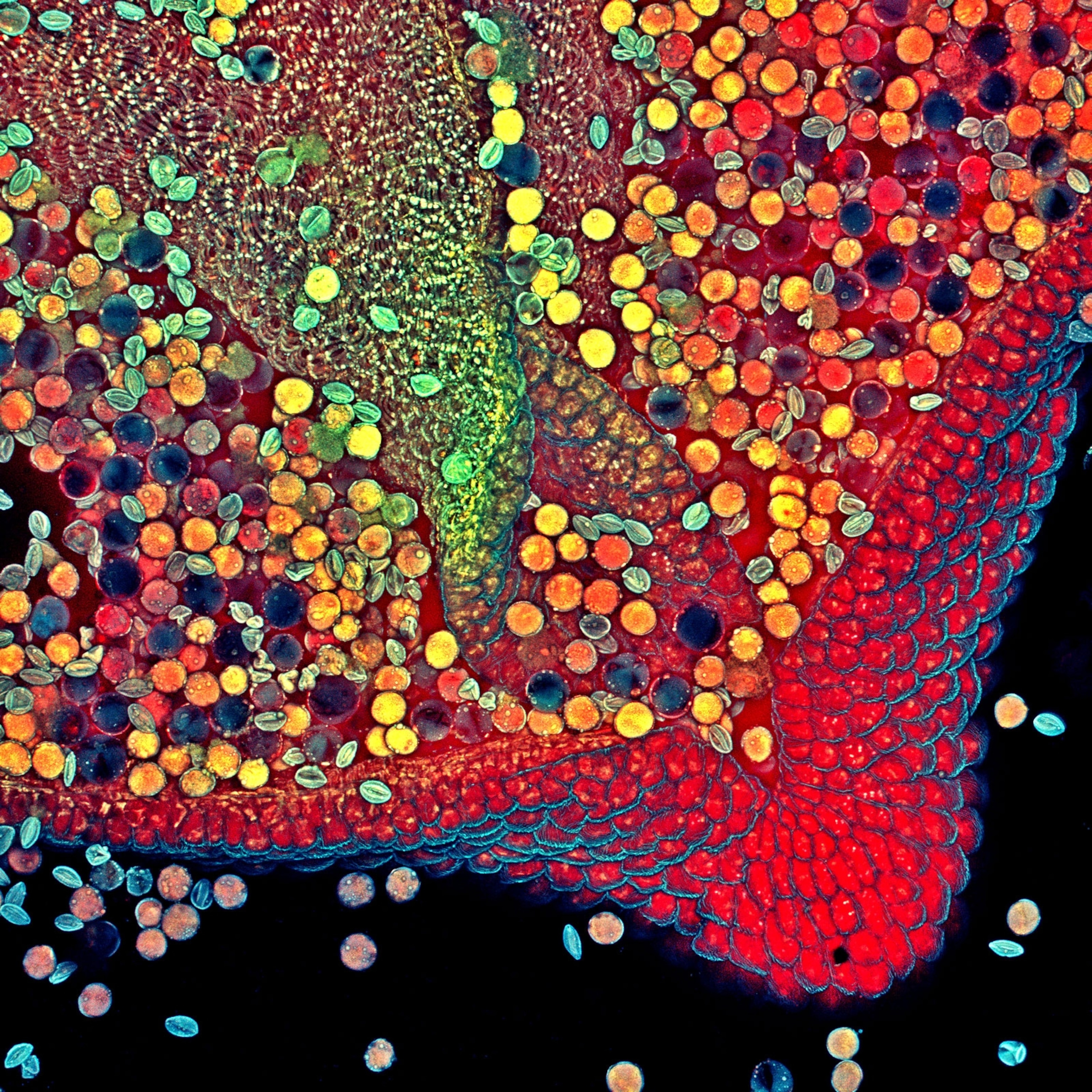
Nikon has announced the winners of its 46th annual Small World Photomicrography Competition.
Researcher Daniel Castranova was about to be kicked off the microscope when he took the picture. A colleague in the Brant Weinstein lab at the National Institutes of Health in Maryland was waiting for a turn on the imaging device, which takes hundreds of frames that later are stitched together. Castranova was bringing a microscopic world into focus, with the hope of revealing unprecedented detail. He had a live juvenile zebrafish suspended in jelly under the lens, and he decided to zoom out from its head for one last shot: a full-body image of the fish. He got it, and then promptly forgot about it. Two weeks later, as he assembled the images, he had a startling result. “I was like, Wow, it came out really nice,” he says.
The ethereal image also happened to represent a groundbreaking find: Zebrafish have lymphatic vessels in their skulls that closely resemble those in mammals and humans. These vessels, which carry fluids essential to the immune system, play a role in a number of neurological diseases, including Alzheimer’s. Because of their speedy reproduction patterns and semitransparency, zebrafish—and those vessels—are easy to study in the lab. The find could lead to a better understanding of the human lymphatic system.
That combination of a beautiful image with significant scientific potential represents the ideal recognized by the Nikon Small World Photomicrography Competition. Now in its 46th year, the competition generated thousands of entries worldwide in 2020, and a judging panel of scientists, photographers, and journalists chose 20 winners, announced today. Castranova’s image took first prize.
“We are proud to showcase imagery that this blend of research, creativity, imaging technology and expertise can bring to scientific discovery,” Nikon said in a statement. “This year’s first place winner is a stunning example.”
Related Topics
You May Also Like


Go Further
Animals
- Octopuses have a lot of secrets. Can you guess 8 of them?
- Animals
- Feature
Octopuses have a lot of secrets. Can you guess 8 of them? - This biologist and her rescue dog help protect bears in the AndesThis biologist and her rescue dog help protect bears in the Andes
- An octopus invited this writer into her tank—and her secret worldAn octopus invited this writer into her tank—and her secret world
- Peace-loving bonobos are more aggressive than we thoughtPeace-loving bonobos are more aggressive than we thought
Environment
- Listen to 30 years of climate change transformed into haunting musicListen to 30 years of climate change transformed into haunting music
- This ancient society tried to stop El Niño—with child sacrificeThis ancient society tried to stop El Niño—with child sacrifice
- U.S. plans to clean its drinking water. What does that mean?U.S. plans to clean its drinking water. What does that mean?
- Food systems: supporting the triangle of food security, Video Story
- Paid Content
Food systems: supporting the triangle of food security - Will we ever solve the mystery of the Mima mounds?Will we ever solve the mystery of the Mima mounds?
History & Culture
- Strange clues in a Maya temple reveal a fiery political dramaStrange clues in a Maya temple reveal a fiery political drama
- How technology is revealing secrets in these ancient scrollsHow technology is revealing secrets in these ancient scrolls
- Pilgrimages aren’t just spiritual anymore. They’re a workout.Pilgrimages aren’t just spiritual anymore. They’re a workout.
- This ancient society tried to stop El Niño—with child sacrificeThis ancient society tried to stop El Niño—with child sacrifice
- This ancient cure was just revived in a lab. Does it work?This ancient cure was just revived in a lab. Does it work?
Science
- The unexpected health benefits of Ozempic and MounjaroThe unexpected health benefits of Ozempic and Mounjaro
- Do you have an inner monologue? Here’s what it reveals about you.Do you have an inner monologue? Here’s what it reveals about you.
- Jupiter’s volcanic moon Io has been erupting for billions of yearsJupiter’s volcanic moon Io has been erupting for billions of years
- This 80-foot-long sea monster was the killer whale of its timeThis 80-foot-long sea monster was the killer whale of its time
Travel
- How nanobreweries are shaking up Portland's beer sceneHow nanobreweries are shaking up Portland's beer scene
- How to plan an epic summer trip to a national parkHow to plan an epic summer trip to a national park
- This town is the Alps' first European Capital of CultureThis town is the Alps' first European Capital of Culture
- This royal city lies in the shadow of Kuala LumpurThis royal city lies in the shadow of Kuala Lumpur
